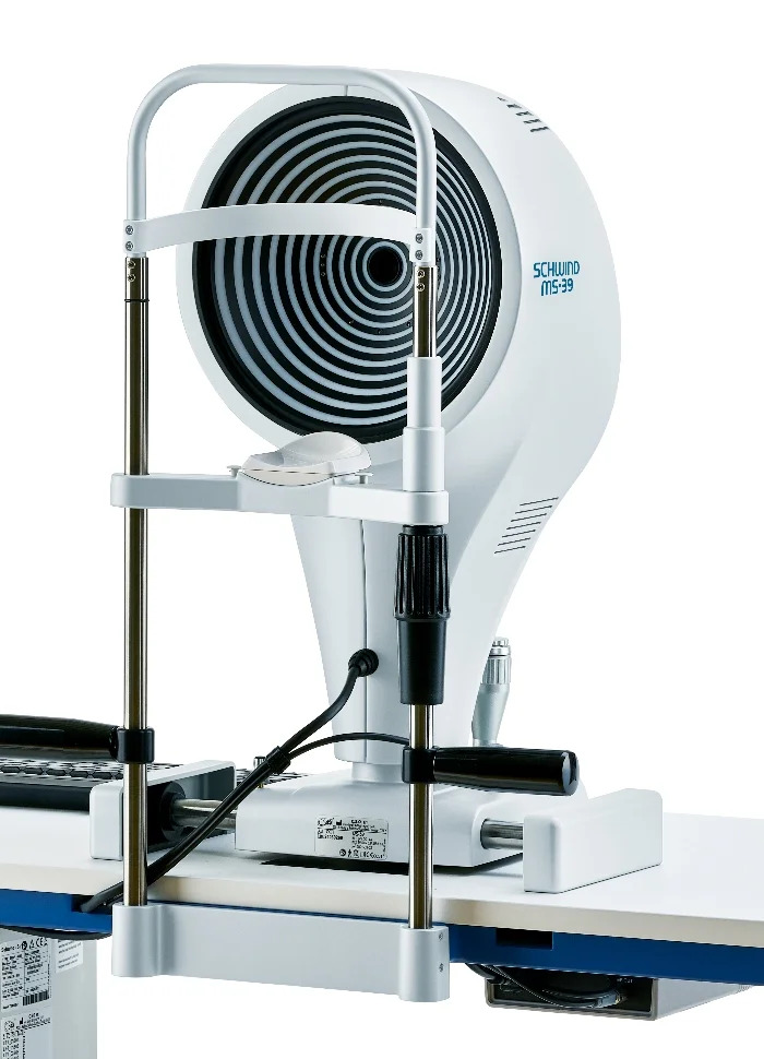
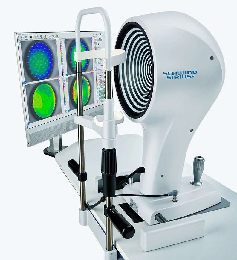
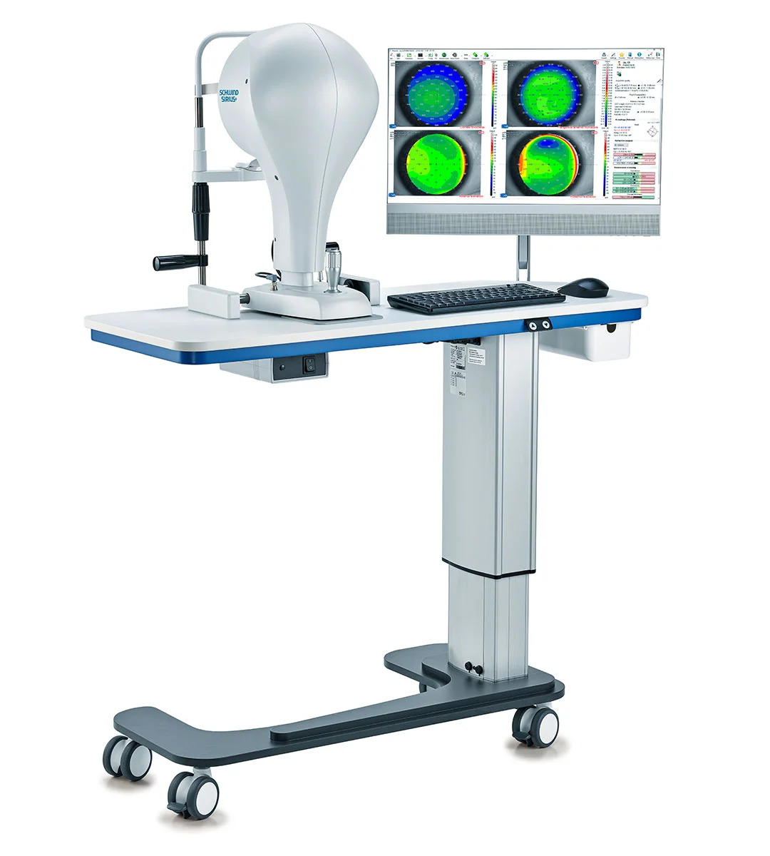
Sirius Tomography in Mumbai
Sirius Tomography stands as a superior technology compared to standard corneal imaging machines due to its advanced utilization of Scheimpflug technology combined with Placido disk topography.
Available at Trivision Eye Hospital in Mumbai, this unique technology captures comprehensive three-dimensional images of the cornea and the anterior segment of the eye. It works by analyzing the refraction of light passing through different layers of the cornea, providing invaluable data for diagnosing conditions like Keratoconus and planning LASIK surgeries.
Personalized Treatment Plans
The detailed information provided by the SIRIUS Tomography allows our ophthalmologists to create highly personalized treatment plans. By tailoring treatments to each individual’s unique corneal characteristics (curvature, thickness, and elevation), the likelihood of achieving optimal refractive outcomes significantly increases.
Enhanced Visual Outcomes
Refractive outcomes heavily depend on the accurate measurement of corneal parameters. The SIRIUS Tomography plays a critical role by providing essential data for precise diagnosis and treatment planning. By leveraging this advanced technology, we can elevate the safety and success rates of our refractive surgeries, ensuring enhanced patient satisfaction with improved visual quality.